
What orthognathic surgery is
It is the surgery that places the upper jaw, the lower jaw or the chin in the right position when the difference between them is too large to correct by moving the teeth alone. It is performed by the maxillofacial surgeon, almost always from inside the mouth, and orthodontics prepares the bite before the operation and finishes it afterwards.
The goal is not only aesthetic: a bite that fits together lets you bite and chew properly, spreads the forces across all the teeth and protects the temporomandibular joint.
When it is indicated
- Skeletal Class III: a protruding lower jaw or a retruded upper jaw; the lower teeth sit in front of the upper ones.
- Skeletal Class II: a retruded lower jaw, with a large gap between the upper and lower front teeth or a profile with a weak chin.
- Skeletal open bite: the front teeth do not meet because of the way the jaws have grown.
- Asymmetries of the jaw and the face.
- Some cases of obstructive sleep apnoea, in which moving the jaws forward widens the airway.
It is considered once the jaws have finished growing. In children, dentofacial orthopaedics uses growth to guide it and, in some cases, avoids surgery or makes it simpler.
How the treatment works, step by step
- 3D study: CBCT, intraoral scan and photographs. We assess the case together with the maxillofacial surgeon and plan the surgery virtually.Before starting, you know what will move and by how much.
- Orthodontics before surgery: we align and coordinate both dental arches so that they fit together once the jaws are in place.With braces or, in selected cases, with aligners. In some cases the surgery can be done at the start.
- Orthognathic surgery: performed in hospital by the maxillofacial surgeon, under general anaesthesia and with a short hospital stay.
- Orthodontics after surgery: a few months to fine-tune the last details of the bite.
- Retention and 3D follow-up: retainers to keep the result and a CBCT superimposition to measure it.
3D planning and follow-up
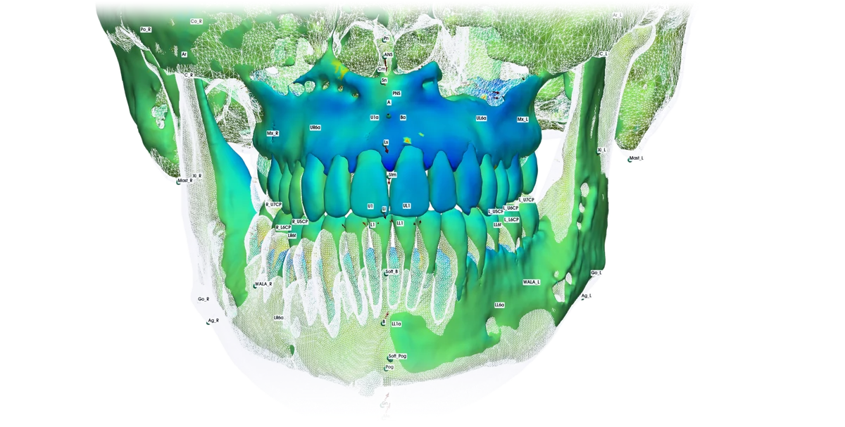
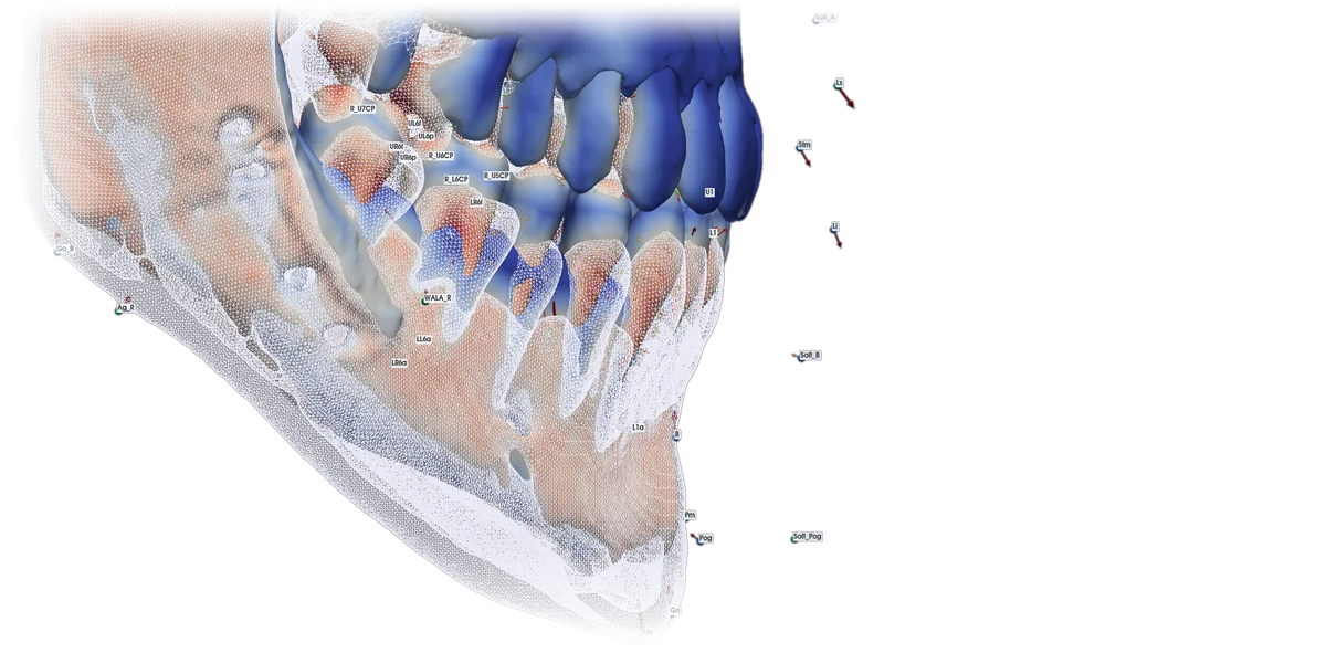
With the CBCT taken before and after, we superimpose both scans in 3D and measure, in millimetres, how much the jaws, the teeth and the chin have moved. The colours show how much each area has changed.
A team effort
The orthodontist and the maxillofacial surgeon decide on the treatment plan together. Dr Manuel Fernández Cano handles the orthodontics from start to finish, coordinates the planning with the surgeon and checks the result in 3D, also taking care of the temporomandibular joint.
The case in the photos
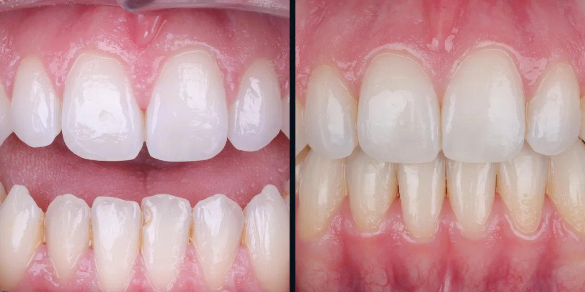
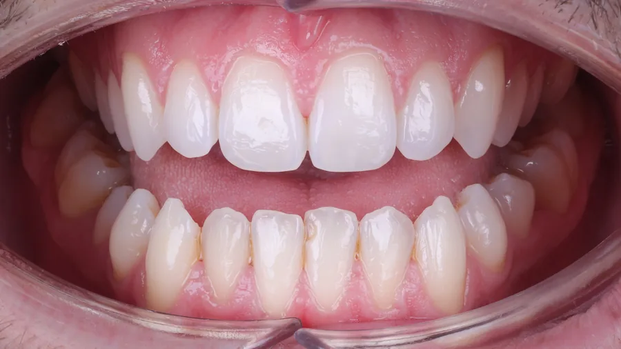
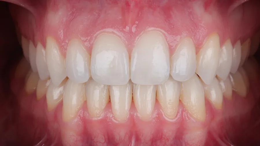
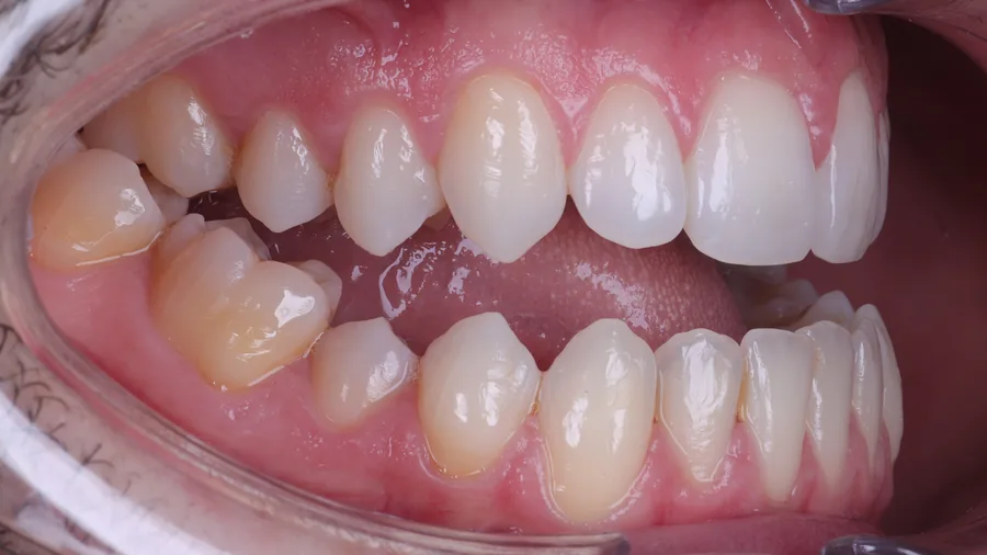
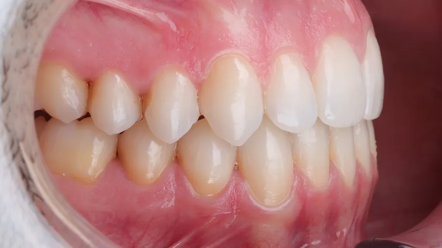
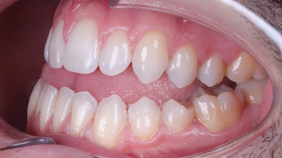
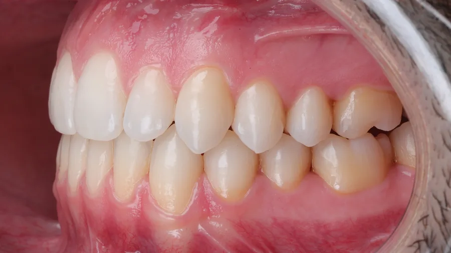
Open bite and Class III: when closing the mouth, the front teeth did not meet. After orthodontics and orthognathic surgery, the incisors fit together and the bite also closes at the sides.
 Before
 After
 Before
 After
 Before
 After
 Book via WhatsApp